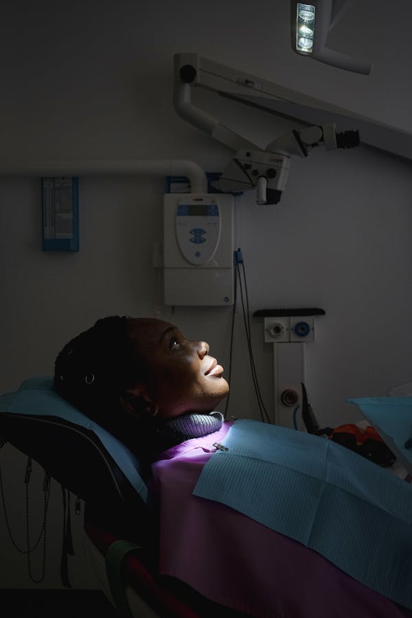
How 3D Scans Made Diagnosis Painless
Goodbye putty impressions — a look inside our fully digital first visit.

The tray of pink putty is history.
For decades, a first dental visit meant biting into a tray of gag-inducing impression material. At Aurora, a wand-sized intraoral scanner captures 3,000 images per second and stitches them into a live 3D model — while you watch it build on screen.
What the scan reveals instantly.
Cracks invisible to the naked eye, early demineralisation between teeth, gum pocket depths and bite imbalances all appear in one model. Your dentist rotates, zooms and measures with you — so the diagnosis is something you see, not something you're told.
Low-dose 3D when roots matter.
Where roots, nerves or implant sites are involved, a cone-beam scan adds the third dimension at a fraction of traditional CT radiation. One 14-second rotation replaces a mouthful of individual X-rays.
What it means for your wallet.
Precision upfront prevents overtreatment later. When both dentist and patient see the same model, plans get smaller, estimates get firmer — and “watch and wait” becomes a confident decision instead of a gamble.